Diffusion MRI maps the diffusion of water molecules in brain tissue. Such information is used by fiber tracking algorithms for tractography of white matter pathways. Below are three examples of studies performed in the context of this workpackage.
Study 1
|
Lyksborg M, Siebner HR, Sørensen PS, Blinkenberg M, Parker GJM, Dogonowski A-M, Garde E, Larsen R, Dyrby TB (2014) Secondary Progressive and Relapsing Remitting Multiple Sclerosis Leads to Motor-Related Decreased Anatomical Connectivity. PLOS ONE 9:e95540. Multiple sclerosis (MS) damages central white matter pathways which has considerable impact on disease-related disability. In this study, diffusion magnetic resonance imaging (dMRI) was used to identify disease-related alterations in anatomical connectivity in patients with relapsing remitting MS (RR-MS) and secondary progressive MS (SP-MS). Anatomical connectivity mapping (ACM) was used to show the connectivity shared between each individual voxel and all other brain voxels. ACM was compared to a commonly used dMRI measure (fractional anisotropy (FA)). In both RR-MS and SP-MS patients, considerable portions of the motor-related white matter revealed decreases in ACM and FA when compared with healthy subjects. Moreover, the results reveal an improved relationship between ACM, the clinical phenotype, and impairment, highlighting the potential of the ACM connectivity indices as a marker for MS. |
 and the corresponding test with FA (b). (c) and (d) show the results of a similar comparison between SP-MS patients and healthy subjects.") |
| The right figure shows where the ACMs of RR-MS patients are decreased relative to healthy subjects (a) and the corresponding test with FA (b). (c) and (d) show the results of a similar comparison between SP-MS patients and healthy subjects. | |
Study 2
Dyrby TB, Lundell H, Burke MW, Reislev NL, Paulson OB, Ptito M, Siebner HR (2014) Interpolation of diffusion weighted imaging datasets. NeuroImage 103:202–213.
Diffusion weighted imaging (DWI) is used to study white-matter fibre organisation, orientation and structural connectivity by means of fibre reconstruction algorithms and tractography. For clinical settings, limited scan time compromises the possibilities to achieve sufficiently high image resolution. We assessed the potential benefits of interpolating DWI datasets to a higher image resolution before fibre reconstruction using a diffusion tensor model. The results indicate that conventional interpolation methods can be successfully applied to DWI datasets for mining anatomical details that are normally seen only at higher resolutions, which will aid in tractography and microstructural mapping of tissue compartments.
 |
 |
|
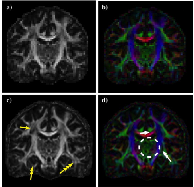
This figure shows the coronal view of a typical DTI reconstructed DWI dataset of the living human brain without a), b) and with c), d) interpolation. a), c) Display of fractional anisotropy values and b), d) with colour-coded directions. Improved contrast to sub structures appears clearly after interpolation to the higher image resolution.
|
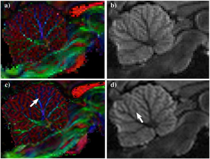
In this figure, the sagittal view of the cerebellum shows that improved anatomical details clearly appear in individual interpolated diffusion weighted images. DWI dataset in original image resolution 0.5 mm3 reconstructed and visualised in a) with colour-coded directions of the tracts, and in b) one of the diffusion weighted image volumes from the DWI dataset. c) and d) show the same as a) and b) after interpolation. Arrows indicate improved geometrical information with interpolation. |
Study 3
Angstmann S, Madsen KS, Skimminge A, Jernigan TL, Baaré WFC, Siebner HR (2016) Microstructural asymmetry of the corticospinal tracts predicts right–left differences in circle drawing skill in right-handed adolescents. Brain Struct Funct:1–15.
 Exploiting structural brain mapping approaches, we pursued the question whether manual dexterity is imprinted in white matter pathways. Whereas most humans show a strong preference to use their right hand, a strong preference for the right hand does not necessarily imply a strong right–left asymmetry in manual proficiency (i.e., dexterity). We tested the hypothesis that intra-individual asymmetry of manual proficiency would be reflected in microstructural differences between the right and left corticospinal tract (CST) in right-handed typically-developing adolescents (11–16 years). Participants were asked to fluently draw superimposed circles with their right dominant and left non-dominant hand. Temporal regularity of circle drawing movements was assessed for each hand using a digitizing tablet. Although all participants were right-handed, there was substantial inter-individual variation regarding the relative right-hand advantage for fluent circle drawing.
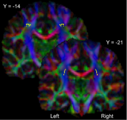
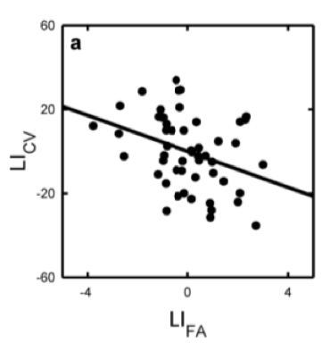 All subjects underwent whole-brain diffusion weighted imaging at 3 Tesla and we applied a tensor model. The right and left CST were defined as regions-of-interest and mean fractional anisotropy (FA) and diffusivity values were calculated for right and left CST. On average, mean FA values were higher in the left CST relative to right CST. The degree of right–left FA asymmetry showed a linear relationship with right–left asymmetry in fluent circle drawing after correction for age and gender.
The higher the mean FA values were in the left dominant CST relative to the right non-dominant CST, the stronger was the relative right-hand advantage for regular circle drawing. This relation was more pronounced in some regions than others but exclusively existent along the CST and not other regions of the white matter skeleton. These findings show that right–left differences in manual proficiency are highly variable in right- handed adolescents and that this variation is associated with a right-left microstructural asymmetry of the CST.