Magnetic resonance: a unique window into the BRAIN
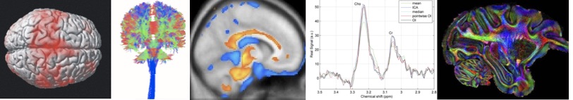
This research area focuses on improving the basic understanding of the brains structure and function by means of novel MR-based imaging and spectroscopic technologies. MRI is unique in the sense that a single scanner modality provides many different imaging contrasts with new and improved sequences continuously coming up. Today, MRI delivers not only exceptional structural information, but also detailed information about function, metabolism and microstructural organization of the tissue. The field moves with an amazing speed where technical barriers are broken almost as fast as they appear and ultrahigh field MRI has become a locomotive for innovation in experimental and clinical research.
We pursue this endeavor by innovating data acquisition techniques but also advancing analytic methods to optimally extract the information from the acquired MR. Our main MRI modalities of interest are diffusion MR, perfusion MR and MR spectroscopy. By keeping in front with the latest MRI technologies and by combining the information with other modalities, such as non-invasive transcranial brain stimulation, we enable high-level basic science and foster research synergies among internal research groups and external collaborators.
MRI: From BENCH to clinic
Our vision is to establish innovative multimodal MRI technologies which offer new insights into brain structure, function, perfusion and metabolic imaging.
We benefit from a unique in-house infrastructure and a cross-disciplinary research environment which allows translational research from the bench to the clinic.
Our translational facilities includes:
- Preclinical laboratories for the creation and working with gen-modified animal models
- An ultra-high field animal 7T Bruker BioSpec MRI scanner with strong gradients, a cryo-coil set-up, and hyperpolarization capabilities for metabolic imaging.
- An ultra-high field 7T Philips MRI scanner for use in humans
- Three clinical 3T scanners (one Philips Achiva scanner, a Siemens Verio scanner, a Siemens Prisma scanner) for brain imaging.
- A well equipped laboratory for hardware design e.g. RF coils