

Intro:
This extensive whole-brain Macaque monkey dataset has been used for validation of structural brain connectivity networks and the impact on them by scanning parameters. The datasets are acquired on an ex vivo monkey brain using diffusion MRI with varying settings. The resulting datasets have different combinations of b-value, gradient angular resolution, and image resolution. The dataset is free for download and use, but please cite our work.
Facts about the dataset:
Sequence: diffusion weighted pulse gradient spin echo (PGSE) with single line readout. Image matrix: 128x256, field-of-view: 64x128 mm2, voxel size: isotropic 0.53 mm3, 91 slices, delta=8ms, DELTA=17ms, TE=30ms. Three shells with different b-values were acquired: b = [1477, 4102, 8040] s/mm² by varying the gradient strength, G = [150, 250, 350] mT/m, TR = [8000, 7900, 8600] ms. Three non-diffusion weighted images (b = 0 s/mm²) were acquired per shell. Each b-value was acquired using a shell with 180 directions, generated by using electrostatic dipole repulsion. The 180 shell was designed to include a subset of 20 and 60 uniformly distributed directions. The protocol was repeated 3 times for the 20 directions protocol and 2 times for the 60 and 180 directions protocols. Lower image resolutions (1.03 and 2.03 mm3) were obtained by linearly down-sampling the 0.53 mm3 images.
Raw dMRI: Datasets with different combinations of the scanning parameters: b-value (1477, 4102, or 8040 s/mm²), angular resolution (20, 60, or 180 directions), and spatial resolution (0.53, 1.03, or 2.03 mm3).
Surfaces: White matter, midcortical, and gray matter surfaces in native diffusion space.
Parcellation: The M132 parcellation containing 91 cortical regions mapped to the b0 image white matter surfaces in native diffusion space.
F99 atlas: T1-weighted image and midcortical surface used to register the M132 parcellation to native diffusion space.
All data have been preprocessed as described in the Ambrosen et al 2020 (Neuroimage) and illustrated in figure 1 also shown below.
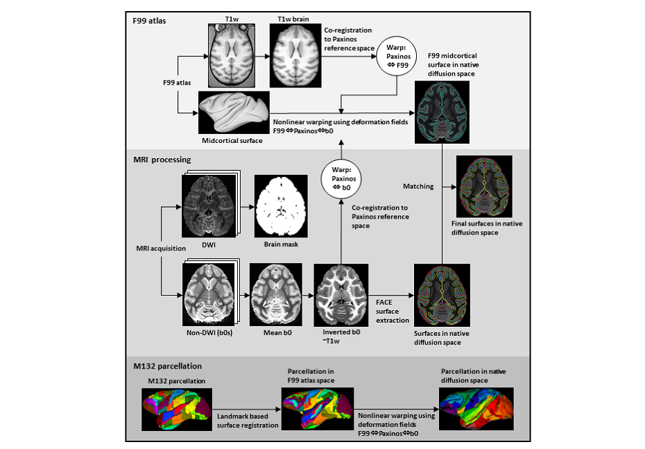
Figure 1 illustrates the pre-processing pipeline to align tracer data, diffusion MRI data and surfaces. The figure is from Ambrosen et al. 2020, NeuroImage, https://doi.org/10.1016/j.neuroimage.2019.116207
Binary Connectivity and Parcellation
The DW-MRI data was used in Girard et al. 2020 in the study of frontal, cingulate, and parietal structural network of the macaque brain. The data was used to build structural connectome and compare the performances of tractography algorithms on the prediction of binary connectivity of 59 cortical areas, well established by the anatomical literature. The 59 areas were manually delineated on the right hemisphere using the average B0 image, following atlas and anatomical literature (see Girard et al 2020).
-Lausanne-Rome-Parcellation.nii.gz: 59-areas cortical parcellation, co-aligned to the DW-MRI data (Figure 2 left).
-tracer-matrix.txt: 59x59 binary connectivity of the macaque brain, derived from anatomical literature (Figure 2 right).
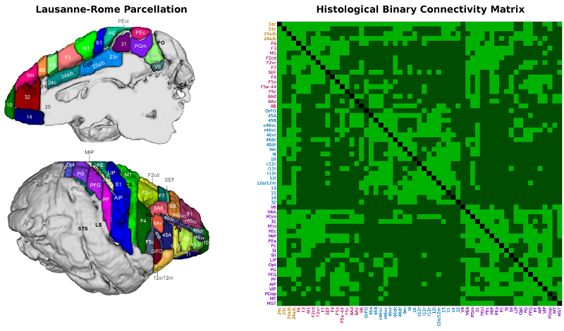
Figure 2 illustrates the 59-areas cortical parcellation (left) and the corresponding binary connectivity obtained from the histological connectivity literature (right).
To cite:
If you use our Rhesus Macaque diffusion MRI data in public or as part of a publication, please cite the following:
Karen S. Ambrosen, Simon F. Eskildsen, Max Hinne, Kristine Krug, Henrik Lundell, Mikkel N. Schmidt, Marcel A.J. van Gerven, Morten Mørup, Tim B. Dyrby, Validation of structural brain connectivity networks: The impact of scanning parameters, (2020) , NeuroImage https://doi.org/10.1016/j.neuroimage.2019.116207
If you use our Laussane-Rome cortical parcellation or binary tracer connectivity matrix, please cite the following:
Gabriel Girard, Roberto Caminiti, Alexandra Battaglia-Mayer, Etienne St-Onge, Karen S. Ambrosen, Simon F. Eskildsen, Kristine Krug, Tim B. Dyrby, Maxime Descoteaux, Jean-Philippe Thiran, Giorgio M. Innocenti, On the cortical connectivity in the macaque brain: a comparison of diffusion tractography and histological tracing data, 2020, NeuroImage.
Download datasets:
Download the diffusion MRI datasets, surfaces, parcellations and templates by clicking HERE.
The Laussane-Rome 59-areas parcellation and the corresponding histological binary connectivity matrix can be download from HERE.
The tracer data used for validation can be downloaded from http://core-nets.org.
Funding:
The Lundbeck Foundation.