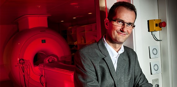
Photo: Joachim Rode
In a few years, small, tiny lesions in the cerebral cortex might be the key to developing precision medicine for patients with sclerosis. Approximately 16,000 people in Denmark – hereof two-thirds women – are diagnosed with the debilitating illness which has frequent symptoms such as motor deprivation, paralysis, sensory deprivation, and impairment of speech.
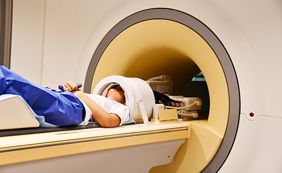
A larger research project at DRCMR, Hvidovre Hospital is currently studying how different lesions in the cerebral cortex, also known as the grey matter of the brain, is connected to the patients’ symptoms – specifically their hand function, which is one of the most frequent symptoms for patients diagnosed with sclerosis. In the project, the most advanced 7 tesla scanner located at the hospital is being used.
"We have decided to make the microscope focus upon the hand area of the brain – meaning the zone in the cerebral cortex which controls our hand movements. We know, in advance, the location of the hands in the cerebral cortex, so now we zoom in and focus upon the details. Thence, we try to construct a map. And later, based upon our observations, we will determine whether the results can be transferred to other functions,” Hartwig Siebner, also Professor in Precision Medicine at University of Copenhagen, explains.
Among others, the project is financed by Skleroseforeningen as well as The Danish Independent Research Fund which most recently has donated almost DKK 2 million.
Fingerprinting in the brain
As a Neurologist, Hartwig Siebner has for years been engaged in the possibility of tailoring the treatment of each individual patient more specifically.
"Using this method, we can develop our understanding of the fingerprints in the brain, set by multiple sclerosis. For some patients, the fingerprints of the disease are found in the cerebral cortex, while they for others mostly are in the white matter of the brain. These are different facets of the same illness,” Hartwig Siebner says.
He believes that in five years it will be possible to separate the diagnoses, so it for some patients can be stated that the white matter plays an important part, while it for other patients is the grey matter which plays the most influential part:
"They will receive slightly different treatments. That way it will be possible to make the medical treatment more accurate,” Hartwig Siebner adds.
The first images of the grey matter
60-80 patients at different stages of their sclerosis are to be scanned – in addition to an equivalent control group without the illness. And then, the MR images are to be compared to specific motor and sensory symptoms – meaning to how the patients are moving and sensing their fingers.
The cerebral cortex – also called cortex or the grey matter – is the just under three-millimetre thin layer located on the exterior part of the white matter of the brain.
"Before we had the 7 tesla scanner, it was practically impossible to get images of lesions in the grey matter. In the white matter, lesions light up more easily. However, it is not until recently that it has become possible for us to focus upon the grey matter, using unique imaging methodology.”
The only scanner of its kind in Denmark
Tesla is a unit of measurement for magnetic field strength. The 7 tesla scanner at Hvidovre Hospital is the only one of its kind in Denmark and is built up around a more than 40 tons heavy electromagnet. The scanner, with the price of more than DKK 40 million, creates a preposterously strong magnetic field, which with the help of the so-called resonance technique makes it possible to get ultra-precise images in high resolution of tiny little areas of the body.
The new images from the grey matter is to be connected with the images of lesions from the white matter, caused by sclerosis – just like the observations are to be connected with many other data – with “immunologically markers, blood tests, and genetics."
Money from external sources
Among others, the project is financed by Skleroseforeningen with DKK 1.9 million, and most recently The Danish Independent Research Fund has donated DKK 1.8 million. The money is to be spent on e.g. the employment of an expert in imaging and data analysis, who is to process an image material of many tera bytes, which is to be thoroughly investigated by a computer:
"One third of the process is to be successful with getting nice, useful images from the scanner. One third is to find a way to process and analyse the images, just making sense of it. And one third is the clinical part – the physiological research – meaning, how do we interpret it. Many factors must be in order – so it is very complex,” Hartwig Siebner states.
Also, an article about the project has been published in Vestegnens Søndagsavis, and you can read it by clicking here.