Tim Dyrby and collaborators have been awarded DKK 4.5 million for three years from the presticious Capital Region Research Foundation for Healthcare as part of its strategic focus on the potential of integrating synchrotron imaging into pre- and clinical Capital Region hospital research.
The world's best X-ray images — seeing nanostructures
Collaboration between researchers from the Capital Region hospitals and the newly opened synchrotron, MAX IV in Lund, Sweden, has opened up for the possibility to make the Capital Region hospitals world-leading in the pre- and clinical use of X-ray imaging with never-seen-before image resolution. By combining the new technique with today's research, X-ray imaging from MAX IV will add a whole new dimension into medical imaging in the understanding of diseases, treatment effects and novel drug designs.
The unique anatomical rich data sets, however, call for new, advanced algorithms to handle and analyse the enormous terabytes of data sets that are acquired during synchrotron experiments, as well as to later visualise the detailed anatomical information arising from these.
The MAXIV Imagers project
The project is led by Tim Dyrby from Hvidovre Hospital and is a synergistic collaboration between several Capital Region hospitals, the University of Copenhagen and the Technical University of Denmark (DTU). The focus is upon the development of new image analyses algorithms that make it possible for both researchers and clinicians to understand the highly detailed anatomical patterns in synchrotron images — and importantly, the potential of the imaging technique.
Synchrotron enables 3D biomedical imaging
We will do four demonstration projects to investigate: 1) the 3D microstructure of the brain, 2) sperm tail movement patterns, 3) leg muscle structure of patients, and 4) bone structure. In doing so, we will probe how to best apply synchrotron imaging in healthcare to make use of the full potential of the technique. The results from the four demonstration projects can be applied directly to clinical projects and thereby pave the way for integrating the potential of synchrotron imaging for improving patient care.
Our vision
We envision that the partners involved in the project will form a leading international competence centre in the pre- and clinical use of synchrotron imaging within biomedical imaging and data analysis. This will provide the Capital Region hospitals with a platform to be internationally pioneering in the use of synchrotron imaging and data analysis.
The MAXIV Imagers Consortium
We will establish a consortium of knowledge for the use of synchrotron imaging for biomedical imaging. The consortium is dynamic and currently includes:
Capital Region hospitals:
- (PI) Associate Professor Tim B. Dyrby, DRCMR, Hvidovre Hospital
- Professor Hartwig R. Siebner, DRCMR. Hvidovre Hospital
- Professor Fin Biering-Sørensen, the NeuroCenter, Rigshospitalet
- Professor Peter Schwartz, Medicinsk Endokrinologisk Klinik, Rigshospitalet
- Senior Researcher Kristian Almstrup, Department of Growth and Reproduction, Juliane Marie Centret, Rigshospitalet
University of Copenhagen:
- Professor Mads Nielsen, DIKU
- Associate Professor Jon Sporring, DIKU
- Postdoc Jessica Pingel, Department of Nutrition, Exercise and Sports
Technical University of Denmark (DTU):
- Professor Rasmus Larsen
- Associate Professor Anders B. Dahl, DTU Compute
International:
- Professor Robert Fiedenhans'l, European XFEL, Germany
- Associate Professor Martin Beck, Lund University/MAX IV, Sweden
- Professor Rajmund Mokso, Lund University, MAX IV/DTU Compute, Sweden
Postdoc Projects:
- Hans Martin Kjer, DTU/DRCMR. 3D reconstruction and data mining of synchrotron data
- Yi He, DRCMR. Translation of synchrotron data of brain tissue to MRI
PhD students in projects:
- Mariam Andersson, DTU Compute/DRCMR. Synchrotron imaging of brain tissue and its relation to microstructural imaging using MRI
- Lise Borg, DIKU. 3D reconstruction of muscle tissue from synchrotron imaging
- Open PhD student position, DIKU/DRCMR. Dynamic tomography reconstruction of synchrotron imaging.
Contact:
What is synchrotron imaging
Just outside the small city of Lund in the Southern part of Sweden, an enormous building has been built (see picture below)
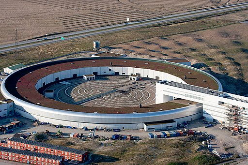
This building houses the national science laboratory, MAX IV, which is a type of facility called a synchrotron, and it consists of two electron rings with circumferences of 528 and 96 meters, respectively.
So, for what is a synchrotron used? The two aforementioned rings contain many electrons that circulate inside. In some areas along the rings, there are magnets that bend the circulating electron beam. When the electron beam is bent, it releases light which we use to study different targets. MAX IV can be thought of as a giant microscope with a light source and beam of higher energy and intensity than any other place in the world.
MAX IV houses the most powerful light source the world has ever seen, and this is what Capital Region researchers have been granted access to! With this light source, molecules and structures smaller than ever seen before can be imaged and investigated. Not only can these structures be imaged — they can be imaged in 3D!
By exposing the target to synchrotron X-ray light from different angles, the 3D structure can be reconstructed. This technique is similar to a medical CT (computed tomography) scan, but with super high resolution that shows structure on a miniscule scale. Advanced visualisation algorithms are needed to display the 3D anatomical fine structure of the sample in question. A combined image of 3-D synchrotron images of muscle fibres and nerve fibres is shown in Figure 1. These 3D visualisations uncover an abundance of information, not previously accessible to researchers.
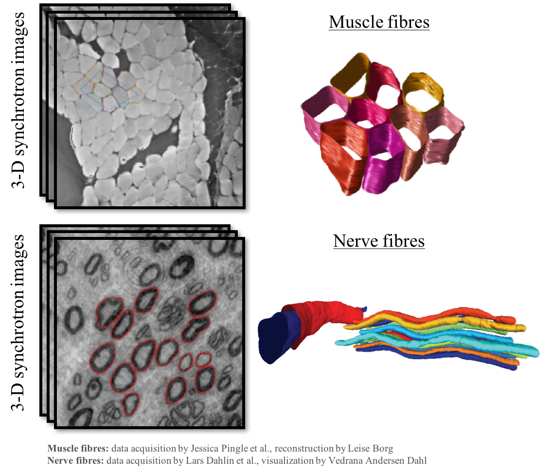
Figure 1