DRCMR Logo 300px Color
 

Quick start guide to the Bloch Simulator

This page provides step-by-step examples to practical use of the Bloch Simulator made for visualizing basic and advanced Magnetic Resonance techniques. The examples are spin-echo formation and the excitation process, which are also demonstrated in corresponding YouTube videos. The basic MR knowledge needed to understand the text below is described in an MRI tutorial targeted at a broad audience.

  • It is easiest to run the simulator directly in an internet browser. If you have not already started it, please do so by following this link starting the software in a new browser window: http://www.drcmr.dk/BlochSimulator
  • If you have Flash installed on your computer, you should now see the magnetization vector of an imaginary sample precessing in a magnetic field, B0, pointing upwards. You can always get back to this situation by choosing "Scene: Precession" in the pull-down menu on the lower left.
  • First try to adjust the B0 field strength by moving the slider named "B0". Notice how the precession frequency changes with the field. Think of the units as tesla, for example.

Spin-echo demonstration, step by step:
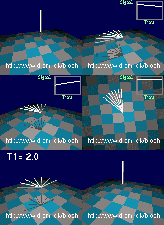
The spin echo sequence
  • Now, let's introduce field inhomogeneity so that all nuclei do not precess at the same frequency. This can be done by choosing "Scene: Weak inhomogeneity" in the pull-down menu on the lower left. You now see the magnetization in thermal equilibrium. The floor rotates since you see this in the rotating frame of resonance which is only needed later. Therefore, shift back to the stationary frame by pressing the  "Change frame"-button.
  • Since weak inhomogeneity was selected above, the nuclei precess at different frequencies, but it does not show until the magnetization is rotated into the transversal plane. Do this by pressing the "90x hard" button (initiates 90 degree RF pulse rotating the magnetization quickly around the x-axis in the transversal plane). You should now see different components of the magnetization precessing at slightly different frequencies. Gradual dephasing leads to a loss of signal seen in the lower right graph. Press the "v" key on keyboard a few times - it toggles the viewing angle.
  • You may want to repeat the last few steps by pressing the "Scene: Weak inhomogeneity" button again and doing a new excitation. Proceed when you understand what you are seeing so far. Let's add a short 180 degree refocusing pulse to our sequence after some dephasing has occurred (press the "180y hard"-button). This will lead to the formation of a spin-echo after a period equal to the dephasing period. Notice how the nuclei get back in phase and how the lost signal is recovered.
  • This finishes the spin-echo demonstration, but you can continue experimenting with it, e.g. by playing out extra refocusing pulses or by adding a bit of relaxation using the T2-slider for example (choose a large value for T2 initially, e.g. 20 seconds). You can also watch the events in the rotating frame of resonance.

Off- and on-resonance excitation, step by step:

  • Go back to the initial situation by  choosing "Scene: Precession" in the pull-down menu on the lower left.
  • Adjust the B0 field strength by moving the slider named "B0". Notice how the precession frequency changes with the field. Set the value of B0 back to 3 by typing the value in the text field above the slider, followed by "enter". Think of the units as tesla, for example.
  • Radio waves applied off-resonance
    Radio waves applied off-resonance
    Now turn on a radio wave field. Do this by setting the RF amplitude to 0.1 using the appropriate slider or by typing the value in the corresponding text field, followed by "enter". The units are the same as for the B0 field. The red bar shows the push of the radio waves on the magnetization (the torque). Notice how the push is orthogonal to the magnetization, and how it rotates.
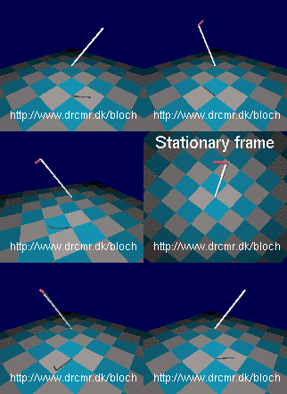
  • The push does not seem to have much of an effect on the magnetization. This is because the frequency of the radio waves is not matched to the Larmor frequency (the radiowaves are off-resonance). The reason for the missing effect may be easier to see in the rotating frame of reference. Press "Change frame" to toggle between viewing in the stationary and the rotating frames of reference as you wish. Notice how the magnetization is not pushed consistently by off-resonance radiowaves (in synchony with the precession). Therefore it just wiggles slightly in the radio wave field. Switch back to the stationary frame of reference.
  • Adjust the radio frequency to match the Larmor frequency simply by setting the RF frequency equal to B0 (3 in this case). This is valid since the gyromagnetic ratio in the simulator is set to 1 Hz/T (approximately). Hence the resonance frequency for a field of value 3 is simply 3. Notice how the magnetization is now gradually changed by the RF field. Watching this in the rotating frame makes it appear much simpler -- switch back and forth a few times, and notice how resonance radiowaves induces rotations in the rotating frame of reference.
  • Radio waves applied on resonance
    Radio waves applied on resonance
    Now you have seen how resonant radiowaves change the magnetization, and how off-resonant waves does not, lets start from equilibrium and do an excitation. Choose "Scene: Equilibrium". Add an RF field by setting the RF amplitude equal to 0.1. Notice how little happens since the RF frequency is not adjusted to the Larmor frequency. Adjust it as described above and see how the magnetization is rotated away from equilibirum, e.g. so it after a while points in the exact opposite directions. Similarly, on-resonance rotations away from equilibrium can be triggered by pressing the buttons labelled "90x hard", "90x selective", "180y hard" and "30x hard" that send bursts of radiowaves sufficiently long to cause the specified rotations.
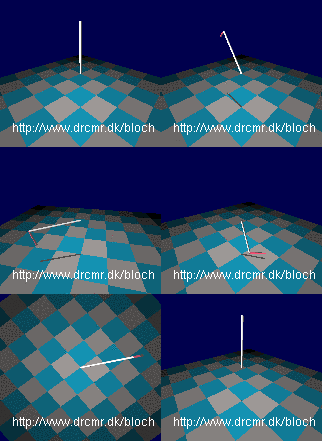

The simulator can do much more. A few examples are given in YouTube videos and on the project homepage but you really need to experiment yourself to get full benefit. The "Challenges" section in the "Help" menu may also be of inspiration.


Selected Publications

Siebner, Thomas Hartwig, Stefan Fuglsang, Christopher Fugl Madelung, Annemette Løkkegaard, Flemming Bendtsen, Jens Dahlgaard Hove, Morten Damgaard, Jan Lysgård Madsen, and Hartwig Roman Siebner. “Gastric Emptying Is Not Delayed and Does Not Correlate With Attenuated Postprandial Blood Flow Increase in Medicated Patients With Early Parkinson’s Disease.” Frontiers in Neurology 13 (2022). https://www.frontiersin.org/article/10.3389/fneur.2022.828069.

Madelung, Christopher F., David Meder, Søren A. Fuglsang, Marta M. Marques, Vincent O. Boer, Kristoffer H. Madsen, Esben T. Petersen, Anne-Mette Hejl, Annemette Løkkegaard, and Hartwig R. Siebner. “Locus Coeruleus Shows a Spatial Pattern of Structural Disintegration in Parkinson’s Disease.” Movement Disorders 37, no. 3 (2022): 479–89. https://doi.org/10.1002/mds.28945.

Siebner, Thomas Hartwig, Christopher Fugl Madelung, Flemming Bendtsen, Annemette Løkkegaard, Jens Dahlgaard Hove, and Hartwig Roman Siebner. “Postprandial Increase in Mesenteric Blood Flow Is Attenuated in Parkinson’s Disease: A Dynamic PC-MRI Study.” Journal of Parkinson’s Disease 11, no. 2 (January 1, 2021): 545–57. https://doi.org/10.3233/JPD-202341.

Herz, Damian M., David Meder, Julia A. Camilleri, Simon B. Eickhoff, and Hartwig R. Siebner. “Brain Motor Network Changes in Parkinson’s Disease: Evidence from Meta-Analytic Modeling.” Movement Disorders 36, no. 5 (2021): 1180–90. https://doi.org/10.1002/mds.28468.

Lohse, Allan, David Meder, Silas Nielsen, Anders Elkjær Lund, Damian M Herz, Annemette Løkkegaard, and Hartwig R Siebner. “Low-Frequency Transcranial Stimulation of Pre-Supplementary Motor Area Alleviates Levodopa-Induced Dyskinesia in Parkinson’s Disease: A Randomized Cross-over Trial.” Brain Communications 2, no. 2 (July 1, 2020): fcaa147. https://doi.org/10.1093/braincomms/fcaa147.

Haagensen, Brian N., Damian M. Herz, David Meder, Kristoffer H. Madsen, Annemette Løkkegaard, and Hartwig R. Siebner. “Linking Brain Activity during Sequential Gambling to Impulse Control in Parkinson’s Disease.” NeuroImage: Clinical 27 (January 1, 2020): 102330. https://doi.org/10.1016/j.nicl.2020.102330.

Betts, Matthew J., Evgeniya Kirilina, Maria C. G. Otaduy, Dimo Ivanov, Julio Acosta-Cabronero, Martina F. Callaghan, Christian Lambert, et al. “Locus Coeruleus Imaging as a Biomarker for Noradrenergic Dysfunction in Neurodegenerative Diseases.” Brain 142, no. 9 (September 1, 2019): 2558–71. https://doi.org/10.1093/brain/awz193.

Meder, David, Damian Marc Herz, James Benedict Rowe, Stéphane Lehéricy, and Hartwig Roman Siebner. “The Role of Dopamine in the Brain - Lessons Learned from Parkinson’s Disease.” NeuroImage, Mapping diseased brains, 190 (April 15, 2019): 79–93. https://doi.org/10.1016/j.neuroimage.2018.11.021.

Irmen, Friederike, Andreas Horn, David Meder, Wolf-Julian Neumann, Philip Plettig, Gerd-Helge Schneider, Hartwig Roman Siebner, and Andrea A. Kühn. “Sensorimotor Subthalamic Stimulation Restores Risk-Reward Trade-off in Parkinson’s Disease.” Movement Disorders 34, no. 3 (2019): 366–76. https://doi.org/10.1002/mds.27576.

Meder, David, and Hartwig Roman Siebner. “Spectral Signatures of Neurodegenerative Diseases: How to Decipher Them?” Brain 141, no. 8 (August 1, 2018): 2241–44. https://doi.org/10.1093/brain/awy195.

Lehericy, Stéphane, David E. Vaillancourt, Klaus Seppi, Oury Monchi, Irena Rektorova, Angelo Antonini, Martin J. McKeown, et al. “The Role of High-Field Magnetic Resonance Imaging in Parkinsonian Disorders: Pushing the Boundaries Forward.” Movement Disorders 32, no. 4 (April 1, 2017): 510–25. https://doi.org/10.1002/mds.26968.

Correia, M. M., Rittman, T., Barnes, C. L., Coyle-Gilchrist, I. T., Ghosh, B., Hughes, L. E. & Rowe, J. B.
Towards accurate and unbiased imaging-based differentiation of Parkinson's disease, progressive supranuclear palsy and corticobasal syndrome.
Brain communications. 2, 1, p. 1-18, fcaa051. 2020.

Kaalund, S. S., Passamonti, L., Allinson, K. S. J., Murley, A. G., Robbins, T. W., Spillantini, M. G. & Rowe, J. B.
Locus coeruleus pathology in progressive supranuclear palsy, and its relation to disease severity.
Acta neuropathologica communications. 8, 1, p. 1-11, 11. 2020.

van der Vegt, J. P. M., Hulme, O. J., Madsen, K. H., Buhmann, C., Bloem, B. R., Münchau, A., Helmich, R. C. & Siebner, H. R.
Dopamine agonist treatment increases sensitivity to gamble outcomes in the hippocampus in de novo Parkinson's disease.
NeuroImage. Clinical. 28, p. 1-8, 102362. 2020.

Dogonowski, A. M., Andersen, K. W., Sellebjerg, F., Schreiber, K., Madsen, K. H. & Siebner, H. R.
Functional neuroimaging of recovery from motor conversion disorder: A case report.
NeuroImage. 190, p. 269-274, 2019.

Dubbioso, R., Manganelli, F., Siebner, H. R. & Di Lazzaro, V.
Fast Intracortical Sensory-Motor Integration: A Window Into the Pathophysiology of Parkinson's Disease.
Frontiers in Human Neuroscience. 13, p. 1-14, 111. 2019.

van Eimeren, T., Antonini, A., Berg, D., Bohnen, N., Ceravolo, R., Drzezga, A., Höglinger, G. U., Higuchi, M., Lehericy, S., Lewis, S., Monchi, O., Nestor, P., Ondrus, M., Pavese, N., Peralta, M. C., Piccini, P., Pineda-Pardo, J. Á., Rektorová, I., Rodríguez-Oroz, M., Rominger, A., Seppi, K., Stoessl, A. J., Tessitore, A., Thobois, S., Kaasinen, V., Wenning, G., Siebner, H. R., Strafella, A. P. & Rowe, J. B.
Neuroimaging biomarkers for clinical trials in atypical parkinsonian disorders: Proposal for a Neuroimaging Biomarker Utility System.
Alzheimer's & Dementia: Diagnosis, Assessment & Disease Monitoring. 11, p. 301-309, 2019.

Siebner, Hartwig R., David Meder, and Damian M. Herz. “FMRI in Parkinson’s Disease.” In FMRI: Basics and Clinical Applications, edited by Stephan Ulmer and Olav Jansen, 417–31. Cham: Springer International Publishing, 2020. https://doi.org/10.1007/978-3-030-41874-8_26

Thomsen, B. L. C., Herz, D. M., Siebner, H. R. & Løkkegaard, A.
Dyskinesier ved Parkinsons sygdom: opdatering om nye billeddannende metoder og behandlingsmuligheder.
Ugeskrift for Laeger. 12, 2017, p. 2-6, 2017.

Lehéricy, S., Vaillancourt, D. E., Seppi, K., Monchi, O., Rektorova, I., Antonini, A., McKeown, M. J., Masellis, M., Berg, D., Rowe, J. B., Lewis, S. J. G., Williams-Gray, C. H., Tessitore, A., Siebner, H. R. & International Parkinson and Movement Disorder Society -Neuroimaging Study Group.
The role of high-field magnetic resonance imaging in parkinsonian disorders: Pushing the boundaries forward.
Movement disorders. 32, 4, p. 510-525, 2017.

Løkkegaard A, Herz DM, Haagensen BN, Lorentzen AK, Eickhoff SB, Siebner HR (2016) Altered sensorimotor activation patterns in idiopathic dystonia - an activation likelihood estimation meta-analysis of functional brain imaging studies. Hum Brain Mapp 37:547-557.

Herz DM,  Haagensen BN, Nielsen SH, Madsen KH, Løkkegaard A, Siebner HR (2016) Resting-state connectivity predicts levodopa-induced dyskinesias in Parkinson’s disease. Mov Disord 31:521-529.

Herz DM, Haagensen BN, Christensen MS, Madsen KH, Rowe JB, Løkkegaard A, Siebner HR (2015) Abnormal dopaminergic modulation of striato-cortical networks underlies levodopa-induced dyskinesias in humans. Brain 138:1658-1666.

Herz DM, Haagensen BN,Christensen MS, Madsen KH, Rowe J, Løkkegaard A, Siebner HR (2014) The acute brain response to levodopa heralds dyskinesias in Parkinson’s disease. Ann Neurol 75:829-836.

Herz DM, Eickhoff SB, Løkkegaard A, Siebner HR (2014) Functional neuroimaging of motor control in Parkinson’s disease: A meta-analysis. Hum Brain Mapp 35:3227-3237. 

van der Vegt JPM, Hulme OJ, Zittel S, Madsen KH, Weiss MM, Buhmann C, Bloem BR, Münchau A, Siebner HR (2013) Attenuated neural response to gamble outcomes in drug-naive patients with Parkinson’s disease. Brain 136:1192-1203.


Group Members

David Meder

Group Leader

Hartwig R. Siebner

Lasse Christiansen

Mattias Rickhag

Show all group members (14)

External Collaborators

Assoc. Prof. Annemette Løkkegaard

Copenhagen University Hospital Bispebjerg


Damian M. Herz, MD, PhD

Nuffield Department of Clinical Neurosciences
University of Oxford


Prof. Stéphane Lehericy, MD, PhD

Institut du Cerveau et de la Moelle épinière – ICM, 
Centre de NeuroImagerie de Recherche – CENIR, Sorbonne Universités, Paris


Prof. James Rowe

Behavioural and Clinical Neuroscience Institute
Dept. of Clinical Neurosciences, University of Cambridge


Prof. Angela Cenci Nilsson

Lund University


Prof. Andrea Kühn,

Charité - Universitätsmedizin Berlin
Movement Disorders and Neuromodulation Section