2021
Albers KJ, Ambrosen KS, Liptrot MG, Dyrby TB, Schmidt MN, Mørup M. 2021. Using connectomics for predictive assessment of brain parcellations. NeuroImage. 238:1-18. https://doi.org/10.1016/j.neuroimage.2021.118170
He Y, Aznar S, Siebner HR, Dyrby TB. 2021. In vivo tensor-valued diffusion MRI of focal demyelination in white and deep grey matter of rodents. NeuroImage. Clinical. 30:1-9. https://doi.org/10.1016/j.nicl.2021.102675
Lundell H, Ingo C, Dyrby TB, Ronen I. 2021. Cytosolic diffusivity and microscopic anisotropy of N-acetyl aspartate in human white matter with diffusion-weighted MRS at 7 T. NMR in Biomedicine. 34(5):1-14. https://doi.org/10.1002/nbm.4304
Perens J, Salinas CG, Skytte JL, Roostalu U, Dahl AB, Dyrby TB, Wichern F, Barkholt P, Vrang N, Jelsing J, Hecksher-Sørensen J. 2021. An Optimized Mouse Brain Atlas for Automated Mapping and Quantification of Neuronal Activity Using iDISCO+ and Light Sheet Fluorescence Microscopy. Neuroinformatics. 19(3):433-446. https://doi.org/10.1007/s12021-020-09490-8
Skoven C, Tomasevic L, Kvitsiani D, Dyrby TB, Siebner HR. 2021. Profiling the transcallosal response of rat motor cortex evoked by contralateral optogenetic stimulation of glutamatergic cortical neurons. bioRxiv. 1-31. https://doi.org/10.1101/2021.04.15.439619
2020
Ambrosen, K. S., Eskildsen, S. F., Hinne, M., Krug, K., Lundell, H., Schmidt, M. N., van Gerven, M. A. J., Mørup, M. & Dyrby, T. B. (2020). Validation of structural brain connectivity networks: The impact of scanning parameters. NeuroImage. 204, p. 1-13, 116207.
Andreasen, S. H., Andersen, K. W., Conde, V., Dyrby, T. B., Puonti, O. T., Kammersgaard, L. P., Madsen, C. G., Madsen, K. H., Poulsen, I. & Siebner, H. R. (2020). Limited Colocalization of microbleeds and microstructural changes after severe traumatic brain injury. Journal of Neurotrauma. 37, 4, p. 581-592.
Cavaliere, C., Aiello, M., Soddu, A., Laureys, S., Reislev, N. L., Ptito, M. & Kupers, R. (2020).
Organization of the commissural fiber system in congenital and late-onset blindness.
NeuroImage. Clinical. 25, 102133.
Nath, V., Schilling, K. G., Parvathaneni, P., Huo, Y., Blaber, J. A., Hainline, A. E., Barakovic, M., Rafael-Patino, J., Frigo, M., Girard, G., Thiran, J-P., Daducci, A., Rowe, M., Rodrigues, P., Prčkovska, V., Aydogan, D. B., Sun, W., Shi, Y., Parker, W. A., Ould Ismail, A. A., Verma, R., Cabeen, R. P., Toga, A. W., Newton, A. T., Wasserthal, J., Neher, P., Maier-Hein, K., Savini, G., Palesi, F., Kaden, E., Wu, Y., He, J., Feng, Y., Paquette, M., Rheault, F., Sidhu, J., Lebel, C., Leemans, A., Descoteaux, M., Dyrby, T. B., Kang, H. & Landman, B. A. (2020). Tractography reproducibility challenge with empirical data (TraCED): The 2017 ISMRM diffusion study group challenge. Journal of magnetic resonance imaging: JMRI. 51, 1, p. 234-249.
Barrett, R. L. C., Dawson, M., Dyrby, T. B., Krug, K., Ptito, M., D'Arceuil, H., Croxson, P. L., Johnson, P., Howells, H., Forkel, S. J., Dell'Acqua, F. & Catani, M. (2020). Differences in frontal network anatomy across primate species. The Journal of Neuroscience. 25 p., 1650-18.
Benavides, F. D., Jin Jo, H., Lundell, H., Edgerton, V. R., Gerasimenko, Y. & Perez, M. A. (2020).
Cortical and Subcortical Effects of Transcutaneous Spinal Cord Stimulation in Humans with Tetraplegia. The Journal of Neuroscience.
Postans, M., Parker, G. D., Lundell, H., Ptito, M., Hamandi, K., Gray, W. P., Aggleton, J. P., Dyrby, T. B., Jones, D. K. & Winter, M. (2020). Uncovering a Role for the Dorsal Hippocampal Commissure in Recognition Memory. Cerebral Cortex. 15 p., bhz143.
Romascano, D., Barakovic, M., Rafael-Patino, J., Dyrby, T. B., Thiran, J-P. & Daducci, A. (2020).
ActiveAxADD: Toward non-parametric and orientationally invariant axon diameter distribution mapping using PGSE. Magnetic Resonance in Medicine.
2019
Bauer, C. (2019). Structural correlates of fatigue in multiple sclerosis assessed with magnetic resonance imaging (MRI). Department of Clinical Medicine, Faculty of Health and Medical Sciences, University of Copenhagen, Copenhagen, Denmark.
Alexander, D. C., Dyrby, T. B., Nilsson, M. & Zhang, H. (2019). Imaging brain microstructure with diffusion MRI: practicality and applications. N M R in Biomedicine. 32, 4, p. 1-26, e3841.
Borg, L., Sporring, J., Dam, E. B., Dahl, V. A., Dyrby, T. B., Feidenhans'l, R., Dahl, A. B. & Pingel, J. (2019). Muscle fibre morphology and microarchitecture in cerebral palsy patients obtained by 3D synchrotron X-ray computed tomography. Computers in Biology and Medicine. 107, p. 265-269.
Innocenti, G. M., Caminiti, R., Rouiller, E. M., Knott, G., Dyrby, T. B., Descoteaux, M. & Thiran, J-P. (2019). Diversity of Cortico-descending Projections: Histological and Diffusion MRI Characterization in the Monkey. Cerebral Cortex. 29, 2, p. 788-801.
Innocenti, G. M., Dyrby, T. B., Girard, G., St-Onge, E., Thiran, J-P., Daducci, A. & Descoteaux, M. (2019). Topological principles and developmental algorithms might refine diffusion tractography. Brain structure & function. 224, 1, p. 1-8.
Lundell, H., Nilsson, M., Dyrby, T. B., Parker, G. J. M., Cristinacce, P. L. H., Zhou, F-L., Topgaard, D. & Lasič, S. (2019). Multidimensional diffusion MRI with spectrally modulated gradients reveals unprecedented microstructural detail. Scientific Reports. 9, 1, p. 1-12, 9026.
Sangari, S., Lundell, H., Kirshblum, S. & Perez, M. A. (2019). Residual Descending Motor Pathways Influence Spasticity after Spinal Cord Injury. Annals of Neurology. 86, 1, p. 28-41.
Schilling, K. G., Nath, V., Hansen, C., Parvathaneni, P., Blaber, J., Gao, Y., Neher, P., Aydogan, D. B., Shi, Y., Ocampo-Pineda, M., Schiavi, S., Daducci, A., Girard, G., Barakovic, M., Rafael-Patino, J., Romascano, D., Rensonnet, G., Pizzolato, M., Bates, A., Fischi, E., Thiran, J-P., Canales-Rodríguez, E. J., Huang, C., Zhu, H., Zhong, L., Cabeen, R., Toga, A. W., Rheault, F., Theaud, G., Houde, J-C., Sidhu, J., Chamberland, M., Westin, C-F., Dyrby, T. B., Verma, R., Rathi, Y., Irfanoglu, M. O., Thomas, C., Pierpaoli, C., Descoteaux, M., Anderson, A. W. & Landman, B. A. (2019). Limits to anatomical accuracy of diffusion tractography using modern approaches.
NeuroImage. 185, p. 1-11.
Lasič, S., Topgaard, D., Nilsson, M. & Lundell, H. (2019). A method of performing diffusion weighted magnetic resonance measurements on a sample. IPC nr. G01R33/56341, G01R33/4835, G01R33/543, G01R33/5608, A61B5/055, Patentnr. 16348580, 9 nov. 2017, Prioritetsdato 9 nov. 2016, Prioritetsnr. SE1651469-7 2019.
Lindhøj, M. B., Henriksen, T., Pedersen, L. & Sporring, J. (2019). Using a high-level parallel programming language for GPU-accelerated tomographic reconstruction. (Accepteret/In press) Using a high-level parallel programming language for GPU-accelerated tomographic reconstruction. p. 27-32.
2018
Lasič, S., Lundell, H., Topgaard, D. & Dyrby, T. B. (2018). Effects of imaging gradients in sequences with varying longitudinal storage time-Case of diffusion exchange imaging. Magnetic Resonance in Medicine. 79, p. 2228-2235.
Nielsen, J. S., Dyrby, T. B. & Lundell, H. (2018). Magnetic resonance temporal diffusion tensor spectroscopy of disordered anisotropic tissue. Scientific Reports. 8, 2930.
Sickmann, H. M., Skoven, C., Bastlund, J. F., Dyrby, T. B., Plath, N., Kohlmeier, K. A. & Kristensen, M. P. (2018). Sleep patterning changes in a prenatal stress model of depression.
Journal of Developmental Origins of Health and Disease. 9, 1, p. 102-111.
Alexander, D. C., Dyrby, T. B., Nilsson, M. & Zhang, H. (2018). Imaging brain microstructure with diffusion MRI: practicality and applications. N M R in Biomedicine.
Dogonowski, A. M., Andersen, K. W., Sellebjerg, F., Schreiber, K., Madsen, K. H. & Siebner, H. R. (2018). Functional neuroimaging of recovery from motor conversion disorder: A case report. NeuroImage.
Dyrby, T. B., Innocenti, G., Bech, M. & Lundell, H. (2018). Validation strategies for the interpretation of microstructure imaging using diffusion MRI. NeuroImage.
Innocenti, G. M., Caminiti, R., Rouiller, E. M., Knott, G., Dyrby, T. B., Descoteaux, M. & Thiran, J-P. (2018). Diversity of Cortico-descending Projections: Histological and Diffusion MRI Characterization in the Monkey. Cerebral Cortex.
2017
Røge, R., Sandø Ambrosen, K. M., Albers, K. J., Eriksen, C. T., Liptrot, M. G., Schmidt, M. N., Madsen, K. H. & Mørup, M. (2017). Whole Brain Functional Connectivity Predicted by Indirect Structural Connections.
Lundell, H. M. H., Ingo, C., Dyrby, T. B. & Ronen, I. (2017). Accurate estimation of intra-axonal diffusivity and anisotropy of NAA in humans at 7T.
Lundell, H. M. H., Nilsson, M., Dyrby, T. B., Parker, G. J. M., Cristinacce, P. L. H., Zhou, F., Topgaard, D. & Lasic, S. (2017). Microscopic anisotropy with spectrally modulated q-space trajectory encoding.
2016
Large I, Bridge H, Ahmed B, Clare S, Kolasinski J, Lam WW, Miller KL, Dyrby TB, Parker AJ, Smith JET, Daubney G, Sallet J Bell AH, Krug K, (2016). Individual differences in the alignment of structural and functional markers of the V5/MT complex in primates, Cerebral Cortex, Accepted.
Donahue C, Sotiropoulos S, Jbabdi S, Hernandez-Fernandez M, Beherens T, Dyrby TB, Kennedy H, Knoblauch K, Coalson T, Glasser M, Van Essen D, (2016). Using Diffusion Tractography to Predict Cortical Connection Strength and Distance: A Quantitative Comparison with Tracers in the Monkey, Journal Neuroscience, 36(25): 6758-6770
Innocenti GM, Dyrby TB, Winther Andersen K, Rouillier EM, Caminiti R (2016). The crossed projection to the striatum in two species of monkey and in humans: Behavioral and evolutionary significance, Cerebral Cortex, doi: 10.1093/cercor/bhw161
2015
Reislev NL, Dyrby TB, Siebner HR, Kupers R, (2015). Simultaneous assessment of white matter changes in microstructure and connectedness in the blind brain, Neural Plasticity, Article ID 795865.
Ratzer R, Iversen P, Börnsen L, Dyrby TB, Christensen JR, Ammitzbøll C, Madsen CG, Garde E, Andersen B, Hyldstrup L, Sørensen PS, Siebner HR, Sellebjerg F, (2015). Monthly oral methylprednisolone pulse treatment in progressive multiple sclerosis, Multiple Sclerosis Journal (accepted).
Shemesh N, Jespersen SN, Alexander DC, Cohen Y, Drobnjak I, Dyrby TB, Finsterbusch J, Koch MA, Kuder T, Laun F, Lawrenz M, Lundell H, Mitra PP, Nilsson M, Özarslan E, Topgaard D, Westin CF, Conventions and nomenclature for double diffusion encoding NMR and MRI, Magn Reson Med, 2015 (accepted)
Barthélemy D, Willerslev-Olsen M, Lundell H, Biering-Sørensen F, Nielsen JB. (2015). Assessment of transmission in specific descending pathways in relation to gait and balance following spinal cord injury, Prog Brain Res. 218:79-101.
Knösche TR, Anwander A, Liptrot MG, Dyrby TB, (2015). Validation of Tractography – Comparison with Manganese Tracing, Human Brain Mapping, Human Brain Mapping, DOI: 10.1002/hbm.22902.
Reislev NL, Kupers R, Siebner HR, Ptito M, Dyrby TB, (2015). Blindness alters the microstructure of the ventral but not the dorsal visual stream. Brain Structure and Function, DOI: 10.1007/s00429-015-1078-8.
Sickmann HM, Arentzen TS, Dyrby TB, PlathN, Kristensen MP, (2015). Prenatal Stress Produces Sex Specific Changes in Depression-like Behavior in Rats: Implications for increased vulnerability in females, Journal of Developmental Origins of Health and Disease, (Accepted).
Lundell H, Sønderby CK, Dyrby TB, (2015). Diffusion weighted imaging with circularly polarized oscillating gradients,Magn Reson Med., 73(3), 171:1176.
Daducci A, Canales-Rodriguez EJ, Zhang H, Dyrby TB, Alexander DC, Thiran JP, (2015). Accelerated Microstructure Imaging via Convex Optimization (AMICO) from diffusion MRI data, Neuroimage, 35, 32-44.
2014
Dyrby TB, Lundell H, Burke MW, Reislev NL, Paulson OB, Ptito M, Siebner HR, (2014). Interpolation of diffusion weighted imaging datasets, NeuroImage, 103, 202–213.
Jespersen SN, Lundel H, Sønderby C, Dyrby TB, (2014). Commentary on ”Microanisotropy imaging: quantification of microscopic diffusion anisotropy and orientation of order parameter by diffusion MRI with magic-angle spinning of the q-vector”, Frontiers in Physics, doi: 10.3389/fphy.2014.00028.
Lundell HM, Alexander DC, Dyrby TB, (2014). High angular resolution diffusion imaging with stimulated echoes: compensation and correction in experiment design and analysis, NMR in Biomedicine.
Liptrot GM, Sidaros K, Dyrby TB, (2014). Addressing The Path-Length-Dependency Confound In White Matter Tract Segmentation, PLOS ONE, 9 (5), e96247.
Lyksborg M, Siebner HR, Sørensen PS, Blinkenberg M, Parker GJM, Dogonowski A, Garde E, Larsen R, Dyrby TB, (2014). Secondary progressive and relapsing remitting multiple sclerosis leads to motor-related decreased anatomical connectivity, PLOS ONE 9 (4), e95540.
Lundell HM, Sønderby CK, Dyrby TB, (2014). Diffusion weighted imaging with circularly polarized oscillating gradients,Magn Reson Med. (accepted).
Christensen JR, Ratzer R, Börnsen L, Lyksborg M, Garde E, Dyrby TB, Siebner HR, Sørensen PS, Sellebjerg F, (2014). Natalizumab in progressive MS – results of an open-label phase 2A proof-of- concept trial. Neurology (accepted).
Sønderby, C. K., Lundell, H. M., Søgaard, L. V. and Dyrby, T. B. (2014). Apparent exchange rate imaging in anisotropic systems. Magn Reson Med., 72(3), 756-762.
2013
Jespersen SN, Lundell H, Sønderby KS, Dyrby TB, (2013). Rotationally invariant sampling of double pulsed field gradient diffusion: estimating apparent compartment eccentricity, NMR in biomedicine, (in press).
Assaf Y, Alexander DC, Jones DK, Bizzi A, Behrens T, Clark C, Cohen Y, Dyrby TB, Huppi P, Knoesche T, LeBihan D, Parker GJM, (2013). CONNECT Consortium, The CONNECT project: Combining Macro- and Micro-structure, NeuroImage, 80, 273-282.
Dyrby TB, Søgaard LV, Hall MG, Ptito M, Alexander DC, (2013). Contrast and stability of the axon diameter index from microstructure imaging with diffusion MRI, Magnetic Resonance in Medicine. 70(3), 711:721.
2012
Lundell H, Barthelemy D, Biering-Sørensen F, Cohen-Adad J, Nielsen JB, Dyrby TB. (2012). Fast diffusion tensor imaging and tractography of the whole cervical spinal cord using point spread function corrected echo planar imaging., Magn Reson Med. 2012 Mar 6. doi: 10.1002/mrm.24235. [Epub ahead of print].
2011
Zhang H, Dyrby TB, Alexander DC., (2011) Axon diameter mapping in crossing fibers with diffusion MRI, Med Image Comput Comput Assist Interv.;14(Pt 2):82-9.
Lundell H, Nielsen J B, Ptito M, Dyrby T B. (2011). Distribution of collateral fibers in the monkey cervical spinal cord detected with diffusion weighted magnetic resonance imaging. NeuroImage, 56(3):923-9.
Dyrby TB, Baaré WFC, Alexander DC, Jelsing J, Garde E, Søgaard LV, (2011). An ex vivo imaging pipeline for producing high-quality and high-resolution diffusion weighted imaging datasets. Hum Brain Mapp. 2011 Apr;32(4):544-63.
2010
Alexander DC, Hubbard PL, Hall MG, Moore EA, Ptito M, Parker GJ, Dyrby TB. Orientationally invariant indices of axon diameter and density from diffusion MRI. Neuroimage. 2010, 52(4):1374-89.
2009
Stavngaard T, Søgaard LV, Batz M, Schreiber LM, Dirksen A. Progression of emphysema evaluated by MRI using hyperpolarized 3He (HP 3He) measurements in patients with alpha-1 antitrypsin deficiency (A1AT), compared with CT and lung function tests. Acta Radiol. 2009, 50(9):1019-26.
2008
Sidaros A, Engberg AW, Sidaros K, Liptrot MG, Herning M, Petersen P, Paulson OB, Jernigan TL, Rostrup E. Diffusion tensor imaging during recovery from severe traumatic brain injury and relation to clinical outcome: a longitudinal study. Brain 2008, 131(Pt 2), 559-572.
2007
Dyrby TB, Sogaard LV, Parker GJ, Alexander DC, Lind NM, Baare WF, Hay-Schmidt A, Eriksen N, Pakkenberg B, Paulson OB, Jelsing J. Validation of in vitro probabilistic tractography. Neuroimage 2007, 37(4), 1267
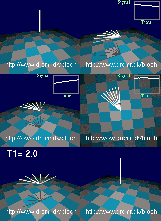
 Radio waves applied off-resonance
 Radio waves applied on resonance


