DRCMR Logo 300px Color
 

Quick start guide to the Bloch Simulator

This page provides step-by-step examples to practical use of the Bloch Simulator made for visualizing basic and advanced Magnetic Resonance techniques. The examples are spin-echo formation and the excitation process, which are also demonstrated in corresponding YouTube videos. The basic MR knowledge needed to understand the text below is described in an MRI tutorial targeted at a broad audience.

  • It is easiest to run the simulator directly in an internet browser. If you have not already started it, please do so by following this link starting the software in a new browser window: http://www.drcmr.dk/BlochSimulator
  • If you have Flash installed on your computer, you should now see the magnetization vector of an imaginary sample precessing in a magnetic field, B0, pointing upwards. You can always get back to this situation by choosing "Scene: Precession" in the pull-down menu on the lower left.
  • First try to adjust the B0 field strength by moving the slider named "B0". Notice how the precession frequency changes with the field. Think of the units as tesla, for example.

Spin-echo demonstration, step by step:
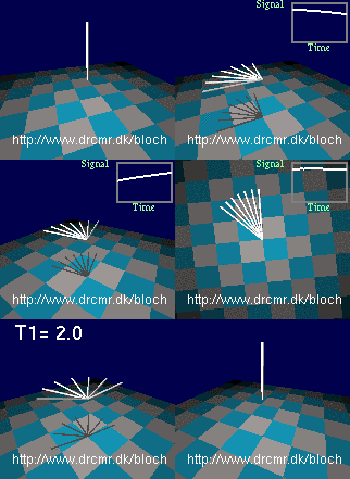
The spin echo sequence
  • Now, let's introduce field inhomogeneity so that all nuclei do not precess at the same frequency. This can be done by choosing "Scene: Weak inhomogeneity" in the pull-down menu on the lower left. You now see the magnetization in thermal equilibrium. The floor rotates since you see this in the rotating frame of resonance which is only needed later. Therefore, shift back to the stationary frame by pressing the  "Change frame"-button.
  • Since weak inhomogeneity was selected above, the nuclei precess at different frequencies, but it does not show until the magnetization is rotated into the transversal plane. Do this by pressing the "90x hard" button (initiates 90 degree RF pulse rotating the magnetization quickly around the x-axis in the transversal plane). You should now see different components of the magnetization precessing at slightly different frequencies. Gradual dephasing leads to a loss of signal seen in the lower right graph. Press the "v" key on keyboard a few times - it toggles the viewing angle.
  • You may want to repeat the last few steps by pressing the "Scene: Weak inhomogeneity" button again and doing a new excitation. Proceed when you understand what you are seeing so far. Let's add a short 180 degree refocusing pulse to our sequence after some dephasing has occurred (press the "180y hard"-button). This will lead to the formation of a spin-echo after a period equal to the dephasing period. Notice how the nuclei get back in phase and how the lost signal is recovered.
  • This finishes the spin-echo demonstration, but you can continue experimenting with it, e.g. by playing out extra refocusing pulses or by adding a bit of relaxation using the T2-slider for example (choose a large value for T2 initially, e.g. 20 seconds). You can also watch the events in the rotating frame of resonance.

Off- and on-resonance excitation, step by step:

  • Go back to the initial situation by  choosing "Scene: Precession" in the pull-down menu on the lower left.
  • Adjust the B0 field strength by moving the slider named "B0". Notice how the precession frequency changes with the field. Set the value of B0 back to 3 by typing the value in the text field above the slider, followed by "enter". Think of the units as tesla, for example.
  • Radio waves applied off-resonance
    Radio waves applied off-resonance
    Now turn on a radio wave field. Do this by setting the RF amplitude to 0.1 using the appropriate slider or by typing the value in the corresponding text field, followed by "enter". The units are the same as for the B0 field. The red bar shows the push of the radio waves on the magnetization (the torque). Notice how the push is orthogonal to the magnetization, and how it rotates.
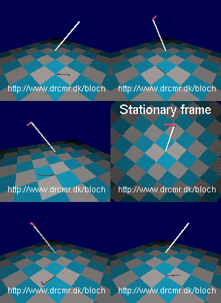
  • The push does not seem to have much of an effect on the magnetization. This is because the frequency of the radio waves is not matched to the Larmor frequency (the radiowaves are off-resonance). The reason for the missing effect may be easier to see in the rotating frame of reference. Press "Change frame" to toggle between viewing in the stationary and the rotating frames of reference as you wish. Notice how the magnetization is not pushed consistently by off-resonance radiowaves (in synchony with the precession). Therefore it just wiggles slightly in the radio wave field. Switch back to the stationary frame of reference.
  • Adjust the radio frequency to match the Larmor frequency simply by setting the RF frequency equal to B0 (3 in this case). This is valid since the gyromagnetic ratio in the simulator is set to 1 Hz/T (approximately). Hence the resonance frequency for a field of value 3 is simply 3. Notice how the magnetization is now gradually changed by the RF field. Watching this in the rotating frame makes it appear much simpler -- switch back and forth a few times, and notice how resonance radiowaves induces rotations in the rotating frame of reference.
  • Radio waves applied on resonance
    Radio waves applied on resonance
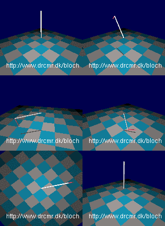
    Now you have seen how resonant radiowaves change the magnetization, and how off-resonant waves does not, lets start from equilibrium and do an excitation. Choose "Scene: Equilibrium". Add an RF field by setting the RF amplitude equal to 0.1. Notice how little happens since the RF frequency is not adjusted to the Larmor frequency. Adjust it as described above and see how the magnetization is rotated away from equilibirum, e.g. so it after a while points in the exact opposite directions. Similarly, on-resonance rotations away from equilibrium can be triggered by pressing the buttons labelled "90x hard", "90x selective", "180y hard" and "30x hard" that send bursts of radiowaves sufficiently long to cause the specified rotations.

The simulator can do much more. A few examples are given in YouTube videos and on the project homepage but you really need to experiment yourself to get full benefit. The "Challenges" section in the "Help" menu may also be of inspiration.


Selected Publications

Dunås T, Wåhlin A, Nyberg L, Boraxbekk C-J. 2021. Multimodal Image Analysis of Apparent Brain Age Identifies Physical Fitness as Predictor of Brain Maintenance. Cerebral Cortex. 31(7):3393-3407. https://doi.org/10.1093/cercor/bhab019

Hansen AL, Boraxbekk C-J, Petersen ET, Paulson OB, Andersen O, Siebner HR, Marsman A. 2021. Do glia provide the link between low-grade systemic inflammation and normal cognitive ageing? A 1H magnetic resonance spectroscopy study at 7 tesla. Journal of Neurochemistry. 159(1):185-196.

Nyberg L, Magnussen F, Lundquist A, Baaré W, Bartrés-Faz D, Bertram L, Boraxbekk CJ, Brandmaier AM, Drevon CA, Ebmeier K, Ghisletta P, Henson RN, Junqué C, Kievit R, Kleemeyer M, Knights E, Kühn S, Lindenberger U, Penninx BWJH, Pudas S, Sørensen Ø, Vaqué-Alcázar L, Walhovd KB, Fjell AM. 2021. Educational attainment does not influence brain aging. Proceedings of the National Academy of Sciences of the United States of America. 118(18):1-3. https://doi.org/10.1073/pnas.2101644118

Sandberg P, Boraxbekk C-J, Zogaj I, Nyberg L. 2021. Ancient Mnemonic in New Format-Episodic Memory Training With the Method of Loci in a Smart Phone Application. Journals of Gerontology - Series B Psychological Sciences and Social Sciences. 76(4):681-691. https://doi.org/10.1093/geronb/gbaa216

Vidal-Pineiro D, Wang Y, Krogsrud SK, Amlien IK, Baaré WF, Bartres-Faz D, Bertram L, Brandmaier AM, Drevon CA, Düzel S, Ebmeier K, Henson RN, Junqué C, Kievit RA, Kühn S, Leonardsen E, Lindenberger U, Madsen KS, Magnussen F, Mowinckel AM, Nyberg L, Roe JM, Segura B, Smith SM, Sørensen Ø, Suri S, Westerhausen R, Zalesky A, Zsoldos E, Walhovd KB, Fjell A. 2021. Individual variations in 'brain age' relate to early-life factors more than to longitudinal brain change. eLife. 10:1-19. https://doi.org/10.7554/eLife.69995

Lind, A., Boraxbekk, C.J., Petersen, E.T., Paulsson, O., Siebner, H., & Marsman, A. (2020). Regional myo-inositol, creatine and choline levels are higher at older age and scale negatively with visuo-spatial working memory: A cross-sectional proton MR spectroscopy study at 7 tesla on normal cognitive ageing. Journal of Neuroscience, 40(42), 8149-8159.

Nyberg, L., Boraxbekk, C.J., Eriksson Sörman, D., Hansson, P., Herlitz, A., Kauppi, K., Ljungberg, J.K., Lövheim, H., Lundquist, A., Nordin Adolfsson, A., Oudin, A., Pudas, S., Rönnlund, M., Stiernstedt, M., Sundström, A., & Adolfsson, R. (2020). Biological and environmental predictors of heterogeneity in neurocognitive ageing: Evidence from Betula and other longitudinal studies. Ageing Research Reviews.

Gylling AT, Bloch-Ibenfeldt M, Eriksen CS, Ziegler AK, Wimmelmann CL, Baekgaard M, Boraxbekk CJ, Siebner HR, Mortensen EL, & Kjaer M. (2020). Maintenance of muscle strength following a one-year resistance training program in older adults. Experimental Gerontology, 139.

Eskilsson, T., Fjellman-Wiklund, A., Ek Malmer, E., Stigsdotter Neely, A., Malmberg Gavelin, H., Slunga Järvholm, L., Boraxbekk, C-J. & Nordin, M.(2020). Hopeful struggling for health: Experiences of participating in computerized cognitive training and aerobic training for persons with stress-related exhaustion disorder. Scandinavian Journal of Psychology.

Friedman, B. B., Suri, S., Solé-Padullés, C., Düzel, S., Drevon, C. A., Baaré, W. F. C., Bartrés-Faz, D., Fjell, A. M., Johansen-Berg, H., Madsen, K. S., Nyberg, L., Penninx, B. W. J. H., Sexton, C., Walhovd, K. B., Zsoldos, E. & Budin-Ljøsne, I. (2020). Are People Ready for Personalized Brain Health? Perspectives of Research Participants in the Lifebrain Consortium. The Gerontologist.

Hansen, A. L., Boraxbekk, C-J., Petersen, E. T., Paulson, O. B., Siebner, H. R. & Marsman, A. (2020). Regional glia-related metabolite levels are higher at older age and scale negatively with visuo-spatial working memory: A cross-sectional proton MR spectroscopy study at 7 tesla on normal cognitive ageing. Cold Spring Harbor Protocols. 36 p., 864496.

Karalija, N., Jonassson, L., Johansson, J., Papenberg, G., Salami, A., Andersson, M., Riklund, K., Nyberg, L. & Boraxbekk, C-J. (2020). High long-term test-retest reliability for extrastriatal 11C-raclopride binding in healthy older adults.
Journal of cerebral blood flow and metabolism.

Wheeler, M. J., Green, D. J., Ellis, K. A., Cerin, E., Heinonen, I., Naylor, L. H., Larsen, R., Wennberg, P., Boraxbekk, C-J., Lewis, J., Eikelis, N., Lautenschlager, N. T., Kingwell, B. A., Lambert, G., Owen, N. & Dunstan, D. W. (2020).
Distinct effects of acute exercise and breaks in sitting on working memory and executive function in older adults: a three-arm, randomised cross-over trial to evaluate the effects of exercise with and without breaks in sitting on cognition.
British Journal of Sports Medicine.

Bangsbo, J., Blackwell, J., Boraxbekk, C-J., Caserotti, P., Dela, F., Evans, A. B., Jespersen, A. P., Gliemann, L., Kramer, A. F., Lundbye-Jensen, J., Mortensen, E. L., Lassen, A. J., Gow, A. J., Harridge, S. D. R., Hellsten, Y., Kjaer, M., Kujala, U. M., Rhodes, R. E., Pike, E. C. J., Skinner, T., Skovgaard, T., Troelsen, J., Tulle, E., Tully, M. A., van Uffelen, J. G. Z. & Viña, J. (2019). Copenhagen Consensus statement 2019: physical activity and ageing. British Journal of Sports Medicine. 53, 14, p. 856-858.

Bojsen-Møller, E., Boraxbekk, C-J., Ekblom, Ö., Blom, V. & Ekblom, M. M. (2019). Relationships between Physical Activity, Sedentary Behaviour and Cognitive Functions in Office Workers.
International Journal of Environmental Research and Public Health. 16, 23, p. 1-15, 4721.

Düzel, E., Acosta-Cabronero, J., Berron, D., Biessels, G. J., Björkman-Burtscher, I., Bottlaender, M., Bowtell, R., Buchem, M. V., Cardenas-Blanco, A., Boumezbeur, F., Chan, D., Clare, S., Costagli, M., de Rochefort, L., Fillmer, A., Gowland, P., Hansson, O., Hendrikse, J., Kraff, O., Ladd, M. E., Ronen, I., Petersen, E., Rowe, J. B., Siebner, H., Stoecker, T., Straub, S., Tosetti, M., Uludag, K., Vignaud, A., Zwanenburg, J. & Speck, O. (2019). European Ultrahigh-Field Imaging Network for Neurodegenerative Diseases (EUFIND). Alzheimer's & dementia (Amsterdam, Netherlands). 11, p. 538-549.

Hedlund, M., Lindelöf, N., Johansson, B., Boraxbekk, C-J. & Rosendahl, E. (2019). Development and Feasibility of a Regulated, Supramaximal High-Intensity Training Program Adapted for Older Individuals. Frontiers in physiology. 10, p. 1-12, 590.

Holm, S. K., Madsen, K. S., Vestergaard, M., Born, A. P., Paulson, O. B., Siebner, H. R., Uldall, P. & Baaré, W. F. C. (2019). Previous glucocorticoid treatment in childhood and adolescence is associated with long-term differences in subcortical grey matter volume and microstructure. NeuroImage. Clinical. 23, p. 1-11, 101825. 

Jonasson, L. S., Nyberg, L., Axelsson, J., Kramer, A. F., Riklund, K. & Boraxbekk, C-J. (2019). Higher striatal D2-receptor availability in aerobically fit older adults but non-selective intervention effects after aerobic versus resistance training. NeuroImage. 202, p. 1-10, 116044.

Magnusson, P. O., Boer, V. O., Marsman, A., Paulson, O. B., Hanson, L. G. & Petersen, E. T. (2019). Gamma-aminobutyric acid edited echo-planar spectroscopic imaging (EPSI) with MEGA-sLASER at 7T.
Magnetic Resonance in Medicine. 81, 2, p. 773-780.

Månsson, K. N. T., Lindqvist, D., Yang, L. L., Svanborg, C., Isung, J., Nilsonne, G., Bergman-Nordgren, L., El Alaoui, S., Hedman-Lagerlöf, E., Kraepelien, M., Högström, J., Andersson, G., Boraxbekk, C-J., Fischer, H., Lavebratt, C., Wolkowitz, O. M. & Furmark, T. (2019). Improvement in indices of cellular protection after psychological treatment for social anxiety disorder. Translational psychiatry. 9, p. 1-10, 340.

Marsman, A. & Pol, H. H. (2019). Glutamaterge neurotransmissie. Handboek schizofreniespectrumstoornissen. 2nd udg. De Tijdstroom/Boom, Bind 2019. p. 370-385..

Malmberg Gavelin, H., Eskilsson, T., Boraxbekk, C.J., Josefsson, M., Stigsdotter Neely, A., & Slunga Järvholm L. (2018). Rehabilitation for improved cognition in patients with stress-related exhaustion disorder: RECO – a randomized clinical trial. Stress.

Boraxbekk, C.-J. (2018). Non-invasive brain stimulation and neuro-enhancement in aging. Clinical Neurophysiology, 129, 464-65.

Baruël Johansen, L., Madsen, K. S., Andersen, K. W., Madsen, K. H., Siebner, H. R. & Baaré, W. F. C. (2017) Reduced orbitofrontal functional network centrality characterizes high neuroticism across childhood and adolescence.

Stomby, A., Otten, J., Ryberg, M., Nyberg, L., Olsson, T. & Boraxbekk, C-J. (2017). A Paleolithic Diet with and without Combined Aerobic and Resistance Exercise Increases Functional Brain Responses and Hippocampal Volume in Subjects with Type 2 Diabetes. Frontiers in Aging Neuroscience. 9, 391.

Jonasson, L. S., Axelsson, J., Riklund, K. & Boraxbekk, C.-J. (2017). Simulating effects of brain atrophy in longitudinal PET imaging with an anthropomorphic brain phantom.
Physics in Medicine and Biology. 62, 13, p. 5213-5227.

Gavelin, H. M., Neely, A. S., Andersson, M., Eskilsson, T., Järvholm, L. S. & Boraxbekk, C-J. (2017). Neural activation in stress-related exhaustion: Cross-sectional observations and interventional effects.
Psychiatry Research. 269, p. 17-25.

Flodin, P., Jonasson, L. S., Riklund, K., Nyberg, L. & Boraxbekk, C.-J. (2017). Does Aerobic Exercise Influence Intrinsic Brain Activity?: An Aerobic Exercise Intervention among Healthy Old Adults.Frontiers in Aging Neuroscience. 9, p. 267.

Eskilsson, T., Slunga Järvholm, L., Malmberg Gavelin, H., Stigsdotter Neely, A. & Boraxbekk, C-J. (2017). Aerobic training for improved memory in patients with stress-related exhaustion: a randomized controlled trial. BMC Psychiatry. 17, 322, p. 1-10, 322.

Bas-Hoogendam, J. M., van Steenbergen, H., Nienke Pannekoek, J., Fouche, J. P., Lochner, C., Hattingh, C. J., Cremers, H. R., Furmark, T., Månsson, K. N. T., Frick, A., Engman, J., Boraxbekk, C.-J., Carlbring, P., Andersson, G., Fredrikson, M., Straube, T., Peterburs, J., Klumpp, H., Phan, K. L., Roelofs, K., Veltman, D. J., van Tol, M. J., Stein, D. J. & van der Wee, N. J. A. (2017). Voxel-based morphometry multi-center mega-analysis of brain structure in social anxiety disorder. NeuroImage: Clinical. 16, p. 678-688.

Jonasson, L.S., Nyberg, L., Kramer, A.F., Lundquist, A., Riklund, K., & Boraxbekk, C.-J. (2017). Aerobic exercise intervention, cognitive performance, and brain structure: Results from the physical influences on brain in aging (PHIBRA) study. Frontiers in Aging Neuroscience; 8, 336.

Boraxbekk, C.-J., Salami, A., Wåhlin, A., & Nyberg, L. (2016). Physical activity over a decade modifies age-related decline in perfusion, gray matter volume, and functional connectivity of the posterior default mode network - a multimodal approach. NeuroImage; 131, 133-141

Group Members

Naiara Demnitz

Group Leader

Sussi Larsen

External Collaborators

Prof. Erik Lykke Mortensen

Department of Public Health, Copenhagen University


Prof. Michael Kjær

Department of Clinical Medicine, Bispebjerg Hospital, University of Copenhagen


Prof. Gunhild Waldemar

Department of Neurology, Copenhagen University Hospital Rigshospitalet 


Prof. Lars Nyberg

Umeå Center for  Functional Brain Imaging