


This free educational MRI and NMR sofware executes on any PC without installation of software. Click to try it now directly in your browser or watch a YouTube introduction to the software. Scroll down for information and software demonstrations.
Background

Magnetic Resonance Imaging (MRI) is a widely used scanning technique based on Nuclear Magnetic Resonance (NMR). When used for medical imaging, the tissue is magnetized by a strong magnetic field present in the scanner. Subsequently the magnetization is manipulated using radio waves and field gradients to make it reflect a physiologically relevant parameter when it is measured. The dynamics are described by the Bloch equations.
It is challenging to learn the basic MR concepts needed for setting up measurements and interpreting results. Fortunately many of the basic aspects can be explored with the Bloch Simulator presented here. It can visualize MR experiments their effect on the magnetization. It has proven very useful for demonstrations in lectures, for student exercises and for self-study.
If you are new to MR, you may first want to read a tutorial and experiment with another piece of software, the JavaCompass, that shows the very basics of Magnetic Resonance using familiar compasses and magnets. When you have also understood the effects of nuclear rotation (spin), causing precession around north rather than vibration through north, you are ready to benefit from the Bloch Simulator presented here.
The Bloch simulator
 The Bloch simulator was written for educational purposes by Lars G. Hanson who got the ESMRMB InfoRESO award for an early version of the software. The simulator is used to explore fundamental aspects of MRI such as precession, resonance, excitation, inhomogeneity and relaxation. Important concepts such as rotating frames, weightings, spoilers, spin-echoes, stimulated echoes and driven equilibrium can also be demonstrated using the program. Finally, the fundamentals of MR imaging can be shown, i.e. how the similarity between induced phase roll patterns and the structure of the imaged object is reflected in the MR signal. Some applications are documented at YouTube, and others here.
The first version of the software was described in RadioGraphics. Since then the graphics and ease of use and installation was improved immensely. A port to Flex/ActionScript was developed by Oddlabs with support from the Danish Ministry of Science, Technology and Innovation.
The simulator is useful during lectures for both technical and non-technical students (radiographers, MDs,...). It has also proven useful for student exercises. Examples are given on this web site and in the software documentation which is distributed with the software (choose "Help" and "Challenges").
Above: Screen dump illustrating the use of the program. Click image one or more times to enlarge. Initial conditions are chosen via menues as shown. Subsequently radio-frequency (RF) pulses can be employed to illustrate MR techniques and sequences. The screen shot is captured during a demonstration of excitation in the presence of field inhomogeneities. The spin isochromates shown in white are pushed by the RF field as indicated by the red bars.
Downloads
 You can run the web version directly from http://www.drcmr.dk/BlochSimulator. This is recommended since the link also provides a "Flash installer" in case software prerequisites are not in place. However, you can also easily download a copy to put on your computer's Desktop, for example. Just right-click on the following link and download ("save to file..." or similar). You can most likely just click on the file subsequently to start the simulator. The now obsolete IDL/Perl implementation is also available for download.
Links to related resources
- Advice on MR teaching, and addditional software, animations and course notes by the author of the Bloch Simulator: http://www.drcmr.dk/MR
- Bloch and imaging simulators: JEMRIS, SIMRI, Odin, SpinBench, MRISIMUL
- MRI e-learning: e-MRI
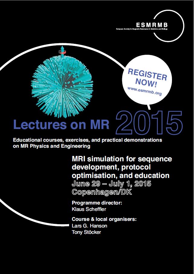
- ESMRMB Lectures on MRI simulation were first given in 2013, and again in June 2015 in Copenhagen. They are likely offered in 2017 again. Venue suggestions are welcome.