DRCMR Logo 300px Color
 

Ultra-High Field MR

The overall focus of the group is to advance the technology and thereby unleash the full potential of ultra-high field MR. Ultra-high field MR offers unique opportunities for revealing new insight into the relationship between structure and function of the human body as part of our clinical research studies.

7T log final


OBJECTIVES

The Ultra-High Field MR group strives at providing state-of-the-art sequences and protocols, which take full advantage of the 7T system available at DRCMR. MR systems operating at fields of 7 tesla or above pose a series of technical challenges for reaching the full potential of the systems:

  • The improved signal-to-noise at 7 tesla allows submillimeter structural and functional image resolution which offers new insights into the understanding of the organization and processes of the body in health and disease. However, a good compensation of subject motion is required to avoid image degradation, which would defeat the purpose of ultra-high resolution imaging. We work on fast image readout approaches and navigator-based correction methods to reduce the effects from motion and the increased physiological noise we unfortunately experience at higher field strength.
  • The higher radiofrequency (RF) at 7 tesla causes strong interactions between the electromagnetic fields and the human body, which makes optimal RF control challenging within the allowed specific absorption rate (SAR) levels using traditional transmit approaches. Our solution is to develop more advanced excitation approaches and to use novel multi-transmit RF technologies available on our state-of-the-art equipment. These new transmit concepts will allow highly improved RF distributions in the human body, and thereby deliver superior image quality at safe SAR levels.
  • Controlling motion and RF requires confinement of inhomogeneities and fluctuations in the main B0 field. We therefore work on advanced shim and dephasing techniques to not only achieve an ideal homogeneous field, but also to take advantage of being able to control the field temporally during the sequences. We exploit this to make novel zoom imaging methods, to exclude unwanted tissue such as fat as well as in advanced RF pulse designs.

RESEARCH PROJECTS

The technical innovations, made by the group, are available and applied to all clinical studies performed on the system. The group’s clinical interest ranges from high-resolution structural, functional and quantitative imaging to advanced spectroscopy editing and imaging. We apply these techniques to aging studies, studies of neurodegenerative diseases, in particular Parkinsonism, neuropsychiatric research and research on various other diseases. Examples of ongoing or upcoming projects typically conducted in synergy with other groups within or outside DRCMR are listed below:

  • Diffusion weighted magnetic resonance spectroscopy at ultra-high field: Unravelling microstructural changes in cerebral white matter in patients with multiple sclerosis Henrik Lundell is currently pursuing the first clinical ultra-high field (7T) MR study in Denmark. In his project, he combines magnetic resonance spectroscopy with diffusion MRI to shed new light into the microstructural alterations in major motor white-matter tract caused by multiple sclerosis. 
  • Brain metabolite changes across the lifespan: a 7T MR study Anouk Marsman and Anna Lind Hansen exploit the high sensitivity of MRS at 7T to look into which role glutamate, GABA, GSH and lactate plays in neurochemical mechanisms that are important in brain development, function and plasticity as well as neuropsychiatric and neurodegenerative diseases.
  • A generalized prospective motion correction framework for improved spectroscopy, structural and angiographic imaging Mads Andersen and Vincent Boer are developing techniques to update the position of the imaging/spectroscopy volume in real-time, as small head motion occur during scanning. Motion is monitored using extra sequence modules (navigators) that acquire dynamic MR data in pauses of the target imaging/spectroscopy sequence.

7T billede til hjemmeside

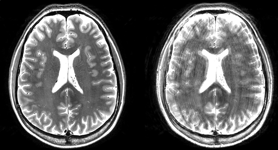
Figure 1: High resolution T2-weighted images acquired at 7T in a subject who moved during the scan. The left image was acquired with motion correction; the right image was acquired without motion correction. The motion was similar in timing and magnitude for the two acquisitions. 

Selected Publications

Marsman A, Lind A, Petersen ET, Andersen M, Boer VO. 2021. Prospective frequency and motion correction for edited 1H magnetic resonance spectroscopy. NeuroImage. 233:1-9. https://doi.org/10.1016/j.neuroimage.2021.117922

Hansen AL, Boraxbekk C-J, Petersen ET, Paulson OB, Andersen O, Siebner HR, Marsman A. 2021. Do glia provide the link between low-grade systemic inflammation and normal cognitive ageing? A 1H magnetic resonance spectroscopy study at 7 tesla. Journal of Neurochemistry. 159(1):185-196.

Lind A, Boraxbekk C-J, Petersen ET, Paulson OB, Siebner HR, Marsman A. 2020. Regional Myo-Inositol, Creatine, and Choline Levels Are Higher at Older Age and Scale Negatively with Visuospatial Working Memory: A Cross-Sectional Proton MR Spectroscopy Study at 7 Tesla on Normal Cognitive Ageing. The Journal of Neuroscience: the official journal of the Society for Neuroscience. 40(42):8149-8159. Available from: 10.1523/JNEUROSCI.2883-19.2020

Boer VO, Andersen M, Lind A, Lee NG, Marsman A, Petersen ET. 2020. MR spectroscopy using static higher order shimming with dynamic linear terms (HOS-DLT) for improved water suppression, interleaved MRS-fMRI, and navigator-based motion correction at 7T. Magnetic Resonance in Medicine. 84(3):1101-1112. Available from: 10.1002/mrm.28202

Magnusson PO, Boer VO, Marsman A, Paulson OB, Hanson LG, Petersen ET. 2019. Gamma-aminobutyric acid edited echo-planar spectroscopic imaging (EPSI) with MEGA-sLASER at 7T. Magnetic Resonance in Medicine. 81(2):773-780. Available from: 10.1002/mrm.27450

Magnusson PO, Boer VO, Marsman A, Paulson OB, Hanson LG, Petersen ET. GABA-edited echo-planar spectroscopic imaging (EPSI) with MEGA-sLASER at 7T. Magn Reson Med, 2018, accepted.

Conference abstracts

ISMRM 2018

Boer VO, Lee NG, Marsman A, Petersen ET. 3D EPSI Hadamard spectral editing of GABA and GSH at 7T. Program no. 1307.

Boer VO, Andersen M, Marsman A, Petersen ET. High resolution imaging at 7T using interleaved prospective motion correction (iMOCO). Program no. 2648.

Lind A, Boer VO, Andersen M, Petersen ET, Marsman A. Test-retest reliability of real-time frequency and motion corrected Hadamard encoded spectral editing (CHASE). Program no. 1291.

Marsman A, Boer VO, Andersen M, Petersen ET. Bilateral functional MRS of GABA with real-time frequency and motion correction at 7T. Program no. 3963.

Nam K, Lee NG, Marsman A, Boer VO, Lee C, Petersen ET. Fast zoomed QSM of the human midbrain at 7T. Program no. 2193.

Piek M, Lee NG, Marsman A, Boer VO, Petersen ET. Investigating the effect of B1 map inaccuracies on advanced pulse design. Program no. 3400.

ISMRM 2017

Andersen M, Boer VO, Marsman A, Petersen ET. A generalized prospective motion corrected framework for improved spectroscopy, structural and angiographic imaging. Program no. 3934.

Boer VO, Lundell H, Dyrby TB, Ronen I, Petersen ET. Dual voxel diffusion weighted MR-spectroscopy.  Program no. 5491

Lee NG, Boer VO, Petersen ET, Cho G. S-ESPIRiT: Estimation of coil sensitivity maps from MR spectroscopic imaging data using ESPIRiT. Program no. 5513.

Magnusson PO, Boer VO, Marsman A, Lundell H, Hanson LG, Petersen ET. GABA-edited echo-planar spectroscopic imaging (EPSI) with MEGA-sLASER at 7T. Program no. 1255.

Marsman A, Boer VO, Andersen M, Petersen ET. Real-time frequency and motion corrected Hadamard encoded spectral editing (CHASE). Program no. 5493.

Zhurbenko V, Boer VO, Petersen ET. Microstrip resonator for high field MRI with capacitor-segmented strip and ground plane. Program no. 4413.

ISMRM 2016

Boer VO, Petersen ET. DC plasma coils for MRI. Program no. 0489.

Zhurbenko V, Boer VO, Petersen ET. Transmission line resonator segmented with series capacitors. Program no. 2160.

Group Members

Vanessa Wiggermann

Group Leader

Henrik Lundell

Group Leader

Michal Povazan

Lars G. Hanson

Hans Stærkind

Jasmin Merhout

Show all group members (11)

External Collaborators

Jeroen Hendrikse

Department of Radiology, University Medical Center Utrecht, The Netherlands


Dennis Klomp

Department of Radiology, University Medical Center Utrecht, The Netherlands


Andrew Webb

Department of Radiology, Leiden University Medical Center, The Netherlands


Matthias van Osch

Department of Radiology, Leiden University Medical Center, The Netherlands


Itamar Ronen

Department of Radiology, Leiden University Medical Center, The Netherlands


Karin Markenroth Bloch

Swedish National 7T facility, Lund, Sweden


Gunther Helms

Swedish National 7T MRI Facility, Medical Radiation Physics, Lund, Sweden


Freddy Ståhlberg

Swedish National 7T MRI Facility, Medical Radiation Physics, Lund, Sweden


Lene Juel Rasmussen

Center for Healthy Aging, University of Copenhagen, Denmark


Erik Lykke Mortensen

Department of Public Health, University of Copenhagen, Denmark


Birte Yding Glenthøj

Center for Neuropsychiatric Schizophrenia Research, Mental Health Services, Capital Region of Denmark, Denmark


Brian Villumsen Broberg

Center for Neuropsychiatric Schizophrenia Research, Mental Health Services, Capital Region of Denmark, Denmark


Kirsten Borup Bojesen

Center for Neuropsychiatric Schizophrenia Research, Mental Health Services, Capital Region of Denmark, Denmark


Egill Rostrup

Functional Imaging Unit, Rigshospitalet Glostrup, Denmark